SIMPLE STAIN TECHNIQUES
|
| Let's suppose we have two bacterial smears containing mixture of Staphylococcus aureus and Escherichia coli.
The organisms are heat fixed by passing an air-dried smear of the organisms through the flame of a gas burner. |
 |
 |
|
| On one of the slides we will apply crystal violet(blue) and on the other one carbol fuchsin or safranin(red). After 1 minute the excess stain is washed off. Allow the smears to air dry. |
|
|
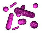
SAFRANIN or CARBOL FUCHSIN |
| Result: |
|
Result: |
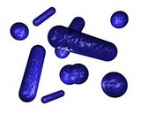 |
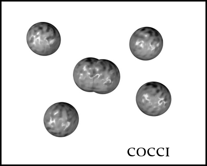
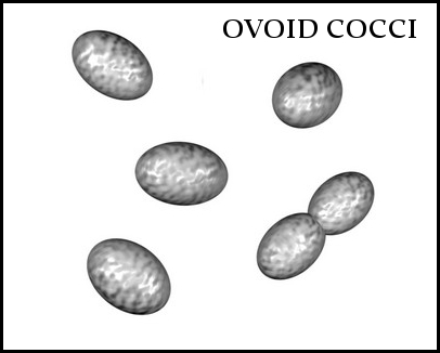
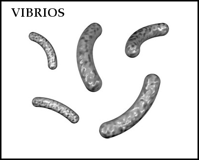
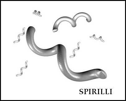
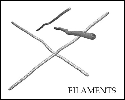
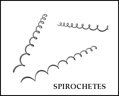
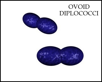
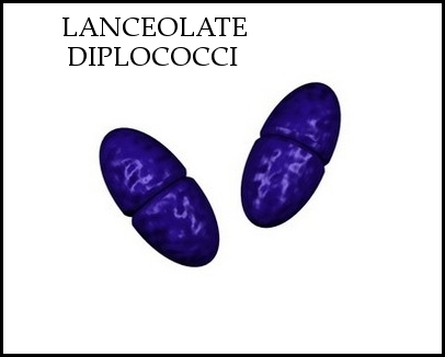
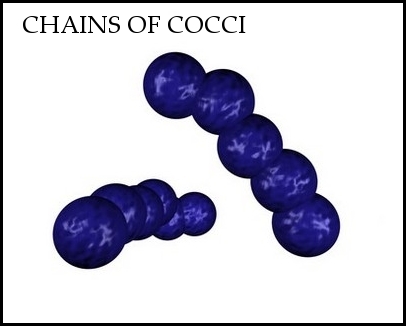
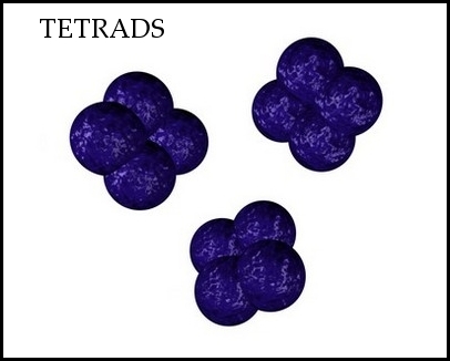
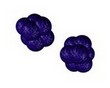
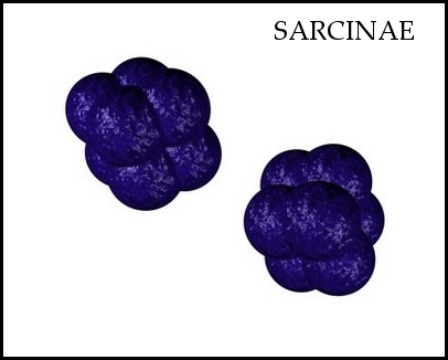
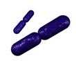
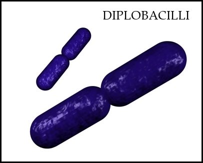
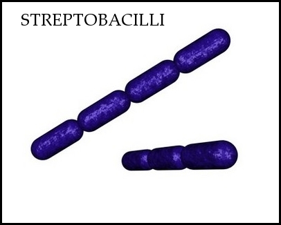
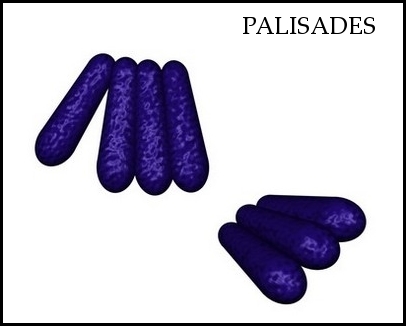
Both staining methods are useful when you want to get some basic information about tested cultures (size, shape, arrangement). |
 |
DIFFERENTIAL STAIN |
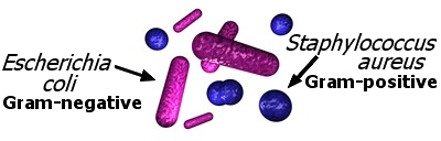
An example is Gram staining (or Gram's method). It is routinely used as an initial procedure in the identification of an unknown bacterial species.
Let's suppose we have a smear containing mixture of Staphylococcus aureus and Escherichia coli as in previous case. We will use the same stains as before and besides we
will need Gram's iodine (strong iodine solution) and alcohol or acetone.
There are four basic steps of the Gram stain:
- applying a primary stain (crystal violet) to a heat-fixed (death by heat) smear of a bacterial culture
- the addition of a trapping agent (Gram's iodine)
- rapid decolorization with alcohol or acetone, and
- counterstaining with safranin or carbol fuchsin (it will more intensely stain anaerobic bacteria)
|
|
Result:
|
Gram staining primarily detects peptidoglycan, which is present in a thick layer in Gram positive bacteria. Gram positive: purple/blue colored cells
Gram negative: pink/red colored cells
|