|
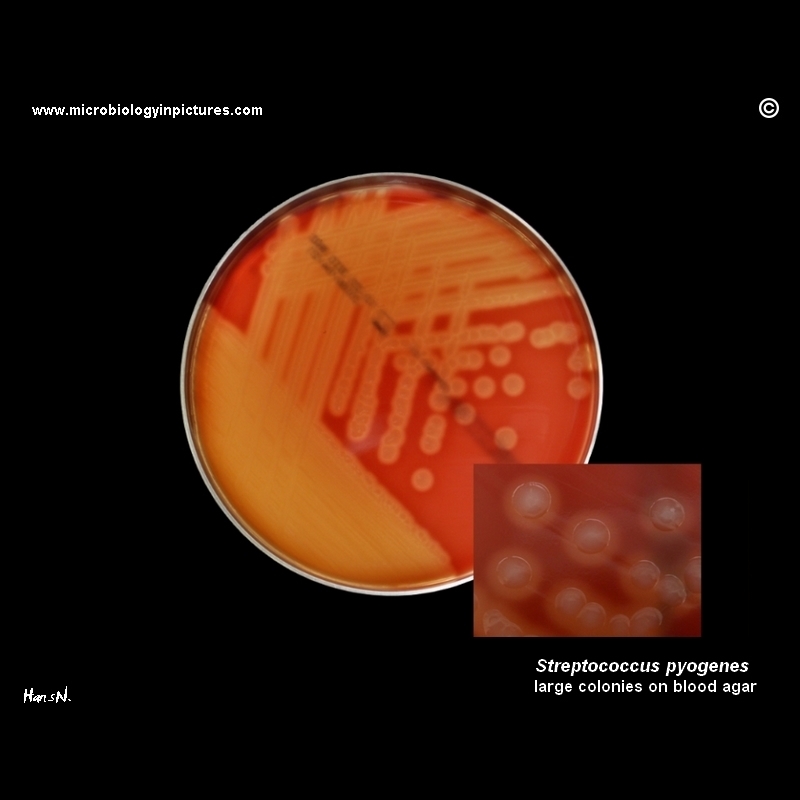
Large colonies of Streptococcus pyogenes cultivated on Columbia agar with 5% sheep blood. Cultivation 24 hours in an aerobic atmosphere enriched with 5% carbon dioxide. Colonies are surroundend by a zone of beta-hemolysis.
Streptococcus pyogenes is the cause of many important human diseases, ranging from mild superficial skin infections to life-threatening systemic diseases. Infections typically begin in the throat or skin. Examples of mild S. pyogenes infections include pharyngitis ("strep throat") and localized skin infection ("impetigo").
Erysipelas and cellulitis are characterized by multiplication and lateral spread of S. pyogenes in deep layers of the skin. S. pyogenes invasion and multiplication in the fascia can lead to necrotizing fasciitis, a potentially life-threatening condition requiring surgical treatment.
|