|
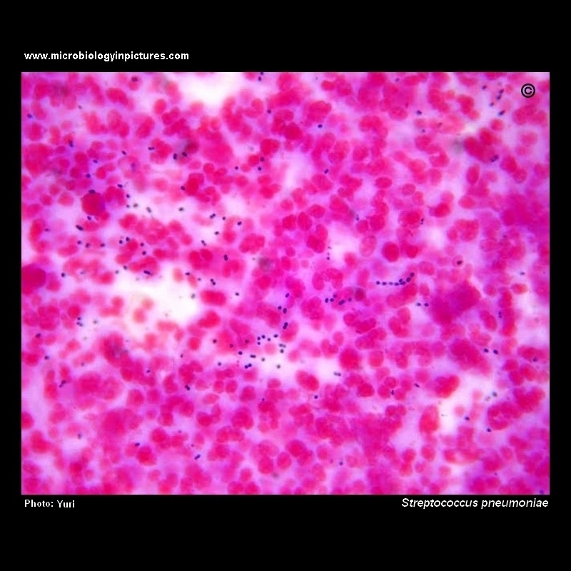
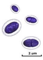
A Gram-stained sample of cerebral spinal fluid (CSF). Pneumococci (Streptococcus pneumoniae) are gram positive cocci and will appear as dark bluish-purple dots in pairs or short chains.
Pneumococci cause 13%19% of all cases of bacterial meningitis in the United States. An estimated 3,000 to 6,000 cases of pneumococcal meningitis occur each year. One-fourth of patients with pneumococcal meningitis also have pneumonia. The clinical symptoms, CSF profile and neurologic complications are similar to other forms of purulent bacterial meningitis. Symptoms may include headache, lethargy, vomiting, irritability, fever, nuchal rigidity, cranial nerve signs, seizures and coma. The case-fatality rate of pneumococcal meningitis is about 30% but may be as high as 80% among elderly persons. Neurologic sequelae are common among survivors. Persons with a cochlear implant appear to be at increased risk of pneumococcal meningitis.
CDCA full story connected with this image you can find on Yuri's Blog |