 |
 |
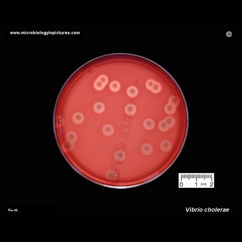
Vibrio cholerae colonies on blood agar |
Hemolytic colonies of Vibrio cholerae on blood agar. Cultivation 24 hours, at a temperature of 37°C.
Vibrio cholerae on blood agar
Hemolytic colonies should have clear zones around them where red blood cells have been totally lysed, and a suspected hemolytic strain should be compared with a strongly hemolytic control strain. Strains that give incomplete hemolysis (incomplete clearing of the red blood cells) should not be reported as hemolytic.
On aerobic sheep blood agar plates, nonhemolytic V. cholerae frequently produces greenish clearing around areas of heavy growth but not around well-isolated colonies. This phenomenon, often describes as hemodigestion, is produced by metabolic by-products which are inhibited by anaerobic incubation of the blood agar plate. For this reason, when aerobic growth conditions are used, hemolysis should be determined around isolated colonies, not in areas of confluent growth. Also, aerobic blood agar plates should be incubated for no more than 18 to 24 hours, since the hemodigestion effect is accentuated during longer incubation periods.
Aerobic incubation of the plate for no longer than 24 hours, although not optimal for detection of hemolysis, will permit differentiation of strongly hemolytic strains, such as the U.S. Gulf Coast and Australia clones, from the nonhemolytic Latin American strains.
|
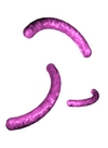
Microscopy:
Gram-negative, curved, non-spore-forming rods. |
|