 |
 |
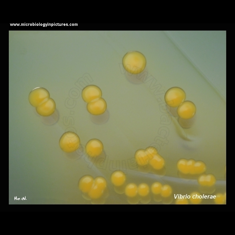
Vibrio cholerae on TCBS |
Colonies of V. cholerae on TCBS agar are large (2-4 mm) and yellow because of the fermentation of sucrose.
TCBS agar is green when prepared. Overnight growth (18 to 24 hours) of V. cholerae will produce large (2 to 4 mm in diameter), slightly flattened, yellow colonies with opaque centers and translucent peripheries. The yellow color is caused by the fermentation of sucrose in the medium. Sucrose nonfermenting organisms, such as V. parahaemolyticus, produce green to blue-green colonies. Suspicious colonies for further testing should be subcultured to a noninhibitory medium, such as gelatin agar, heart infusion agar (HIA), Kliglers iron agar (KIA), or triple sugar iron agar (TSI).
|
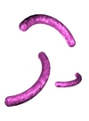
Microscopy:
Gram-negative, curved, non-spore-forming rods. |
|