|
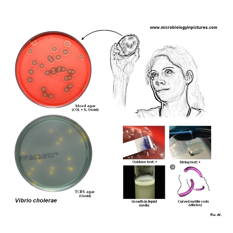
Hemolysis on blood agar
Historically, the classical and El Tor biotypes were differentiated by the ability of the El Tor group to lyse erythrocytes. However, by 1972 almost all El Tor isolates worldwide were nonhemolytic.
The two exceptions to this trend have been the U.S. Gulf Coast and the Australia clones of V. cholerae O1, which are strongly hemolytic when assayed by either the plate or tube hemolysis assay. For this reason, hemolysis continues to be a useful phenotypic characteristic for differentiating the Gulf Coast and Australia clones of V.cholerae O1 from El Tor strains from the rest of the world, including Latin America.
Thiosulfate citrate bile salts sucrose agar (TCBS)
TCBS is the medium of choice for the isolation of V. cholerae and is widely used worldwide. TCBS agar is green when prepared. Sodium citrate, sodium thiosulfate, sodium cholate, and Oxbile are selective agents, providing an alkaline pH to inhibit Gram-positive organisms and suppress coliforms. An increased pH is used to enhance growth of Vibrio cholerae, because this organism is sensitive to acid environments.Overnight growth (18 to 24 hours) of V. cholerae will produce large (2 to 4 mm in diameter), slightly
flattened, yellow colonies with opaque centers and translucent peripheries . The yellow color is caused by the fermentation of sucrose in the medium. Sucrose nonfermenting organisms, such as V. parahaemolyticus, produce green to blue-green colonies.
Oxidase test
Do not use growth from thiosulfate citrate bile salts sucrose (TCBS) agar. Conduct the oxidase test with fresh growth from an HIA slant or any non-carbohydrate-containing medium.
Positive and negative controls should be tested at the same time. Organisms of the genera Vibrio, Neisseria, Campylobacter, Aeromonas, Plesiomonas, Pseudomonas, and Alcaligenes are all oxidase positive; all Enterobacteriaceae are oxidase negative.
String test
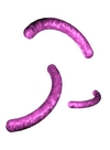
The string test may be performed on a glass microscope slide or plastic petri dish by suspending 18- to 24-hour growth from HIA or other noninhibitory medium in a drop of 0.5% aqueous solution of sodium deoxycholate. If the result is positive, the bacterial cells will be lysed by the sodium deoxycholate, the suspension will lose turbidity, and DNA will be released from the lysed cells causing the mixture to become viscous. A mucoid string is formed when an inoculating loop is drawn slowly away from the suspension. Most vibrios are positive, whereas Aeromonas strains are usually negative.
|